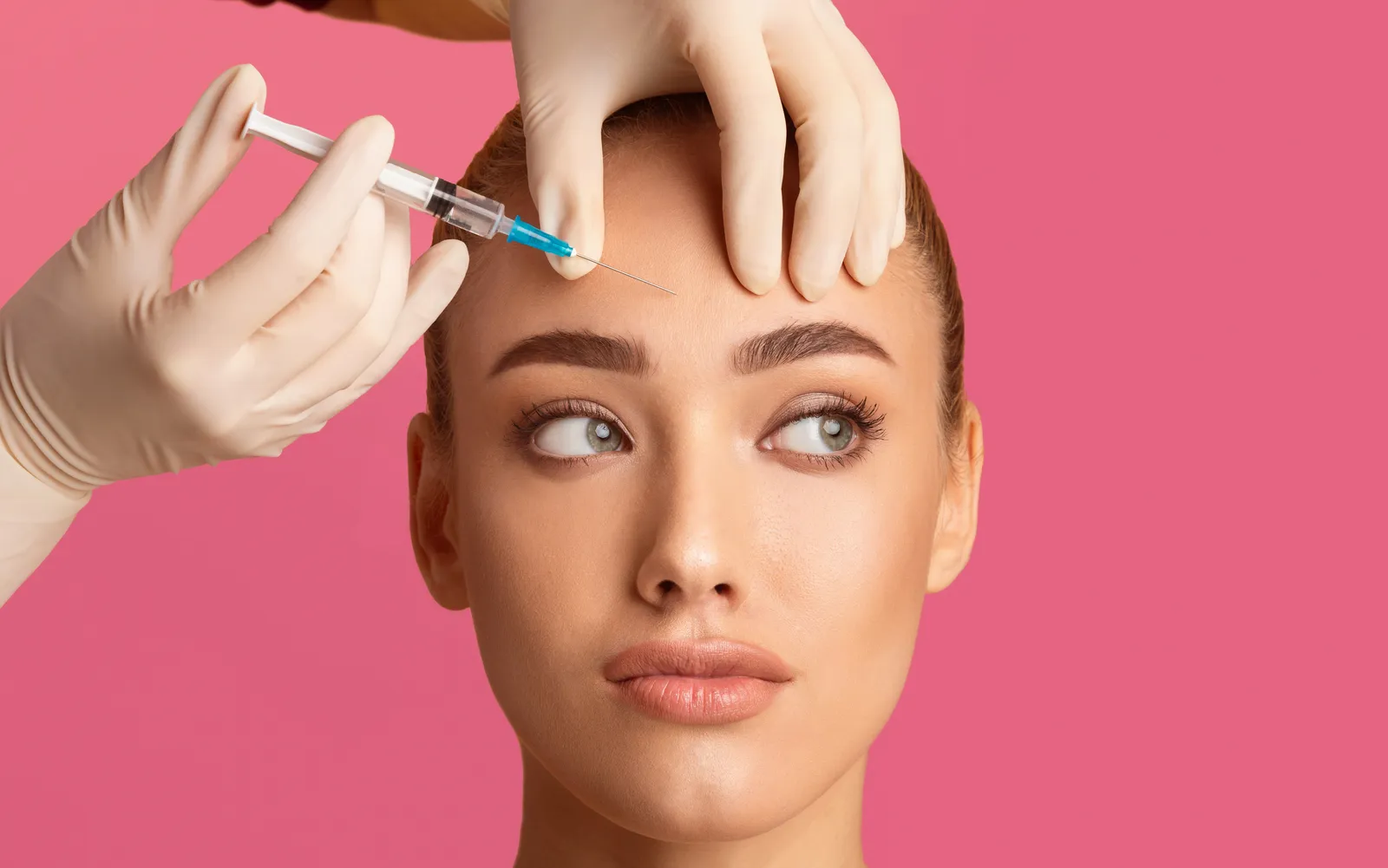

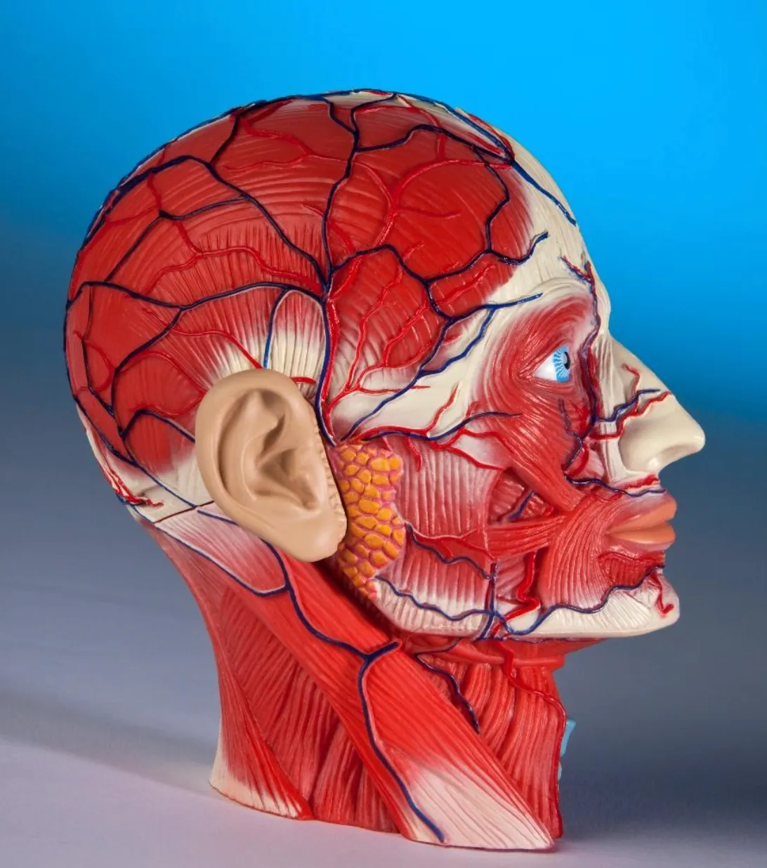
Great aesthetic outcomes begin with anatomy—not the syringe.
Aesthetic medicine has evolved dramatically. We have more injectable products, energy-based devices, and regenerative treatments than ever before. Yet one principle remains unchanged: the most important tool an aesthetic injector brings to the treatment room is a thorough understanding of facial anatomy.
Understanding anatomy is not simply memorizing the names of muscles, arteries, and fat compartments. It means appreciating how these structures interact, how they vary from patient to patient, how aging changes their relationships, and how an intervention in one area can affect the appearance or function of another.
For aesthetic healthcare professionals, anatomical knowledge is the foundation of sound clinical judgment, natural-looking results, and patient safety.
Here are 10 essential principles every aesthetic injector should understand and continually revisit.
1. The Face Is a Multilayered, Integrated Structure
Treating the visible fold alone may not address the underlying anatomy responsible for its appearance.
Similarly, a hollow temple is not simply a volume deficit. Its appearance reflects the relationship between skin, fat, fascia, temporalis muscle, and underlying bone.
Clinical pearl: Before selecting a treatment, identify the anatomical structures contributing to the concern. The most visible feature is not always the primary problem.
2. Facial Vascular Anatomy Is Fundamental to Injection Safety
Vascular anatomy is among the most important subjects an injector must master.
The facial arterial system includes branches of the facial artery, ophthalmic artery, and other vessels that communicate through an extensive network of anastomoses. Important vessels include the angular, facial, supratrochlear, supraorbital, dorsal nasal, and infraorbital arteries, among others.
Their courses, depths, branching patterns, and relationships to surrounding structures vary considerably between individuals.
This variability has important clinical implications. A vessel may be superficial in one patient and deeper in another. A familiar anatomical landmark does not guarantee that a vessel will follow a textbook course.
Vascular compromise following injectable filler treatment can result in skin ischemia, tissue necrosis, and, in rare cases, permanent visual impairment or blindness. The consequences can be devastating, making prevention, early recognition, and prompt management essential.
Every injector should understand:
- The major arterial territories of the face and their potential interconnections.
- How vascular anatomy varies across facial regions and between individuals.
- The clinical signs of vascular compromise, including unexpected pain, blanching, livedoid discoloration, and delayed capillary refill.
- The need for an established emergency protocol, appropriate training, and immediate access to indicated rescue measures.
It is equally important to understand that aspiration, cannula use, and selection of a particular injection plane do not eliminate vascular risk.
Clinical pearl: Never assume that a facial region is completely safe because it is familiar or frequently treated. Anatomical knowledge reduces risk; it does not abolish it.
3. Facial Muscles Must Be Understood Dynamically

Neuromodulator treatment changes muscle activity, and its aesthetic effect depends on the balance between muscles that elevate, depress, or otherwise move a region.
For example, the frontalis is an important eyebrow elevator. Weakening it in a patient who relies heavily on frontalis activity to maintain eyebrow position can contribute to brow heaviness or ptosis-like changes in appearance.
The goal is not simply to reduce movement. It is to understand which movement the patient wants to modify while preserving the expressions and functions that matter to them.
Clinical pearl: Evaluate the face both at rest and in motion. A static photograph cannot fully explain dynamic facial anatomy.
4. Nerves Matter as Much as Muscles
The facial nerve, cranial nerve VII, provides motor innervation to the muscles of facial expression. Its branches include the temporal, zygomatic, buccal, marginal mandibular, and cervical branches, with anatomical variation and interconnections between branches.
The trigeminal nerve, cranial nerve V, provides most facial sensation through its ophthalmic, maxillary, and mandibular divisions.
These systems serve different functions, but both are relevant to aesthetic practice. Motor nerve anatomy helps explain facial movement and the potential consequences of nerve injury or dysfunction. Sensory anatomy is important when evaluating pain, altered sensation, and treatment-related symptoms.
The course of facial nerve branches is particularly relevant when considering procedures involving the temples, lateral face, mandibular border, and other regions where nerve branches may be vulnerable.
Injectors should also recognize pre-existing asymmetry or weakness. A patient with an underlying facial nerve disorder may require a substantially different assessment and treatment plan from someone with uncomplicated age-related changes.
Clinical pearl: Document baseline facial movement and symmetry before treatment. New weakness or asymmetry should never automatically be attributed to a routine aesthetic response.
5. Fat Compartments and Retaining Ligaments Shape Facial Contours
Facial fat is organized into superficial and deep compartments rather than forming one uniform layer.
These compartments contribute differently to facial projection, transitions between anatomical regions, and the appearance of aging. Their size, distribution, and relationship to adjacent structures vary among individuals.
Retaining ligaments provide important connections between deeper tissues and more superficial structures. Together with the SMAS, fat compartments, skin, and underlying skeleton, they influence how the face is supported and how its contours change over time.
As patients age, some fat compartments may lose volume, while others may enlarge or descend. Ligamentous attachments and surrounding tissues also change, producing transitions such as midface flattening, deeper folds, jowling, and changes in the jawline.
This explains why adding volume to every hollow or fold is not always the best approach. Excessive or poorly planned volume can obscure natural contours, create heaviness, or disrupt the balance between adjacent facial regions.
A thoughtful assessment considers whether the patient's concern is primarily related to volume loss, tissue descent, skeletal support, muscle activity, skin quality, or a combination of these factors.
Clinical pearl: Restore balance rather than simply filling depressions. The objective is to improve the relationship between facial structures, not maximize volume.
6. Facial Anatomy Changes With Aging

For example, changes in the maxilla and surrounding midface can affect support of overlying soft tissue. Changes in the mandible can influence lower-face projection and the appearance of the jawline. Meanwhile, dermal thinning and reduced elasticity can make surface irregularities more apparent even when deeper volume remains relatively preserved.
This is why a treatment plan designed for a younger patient with isolated volume loss may be inappropriate for an older patient whose concern reflects several interacting anatomical changes.
Clinical pearl: Treat the patient's anatomy as it exists today—not the anatomy you expect based on age alone.
7. The Periorbital Region Requires Exceptional Anatomical Respect
The periorbital region is among the most anatomically complex areas of the face. It contains delicate skin, the orbicularis oculi, specialized eyelid structures, important neurovascular anatomy, and close relationships with the orbit and ocular circulation.
Aesthetic concerns in this region can include tear-trough hollowing, infraorbital shadowing, dynamic rhytides, skin laxity, and changes in the position or prominence of surrounding tissues.
These concerns can look similar but have different anatomical causes.
For example, infraorbital darkness may result from pigmentation, visible vasculature, shadowing from contour changes, or a combination of factors. Adding volume will not necessarily correct every cause and may worsen edema or produce persistent contour irregularities in an unsuitable patient.
Periorbital treatments also require an appreciation of the potential consequences of vascular compromise. The ophthalmic circulation and its connections with facial vessels make this region particularly important when considering filler-related risk.
Clinical pearl: Distinguish pigmentation, shadowing, volume loss, and edema before recommending treatment. Not every tear trough should be filled.
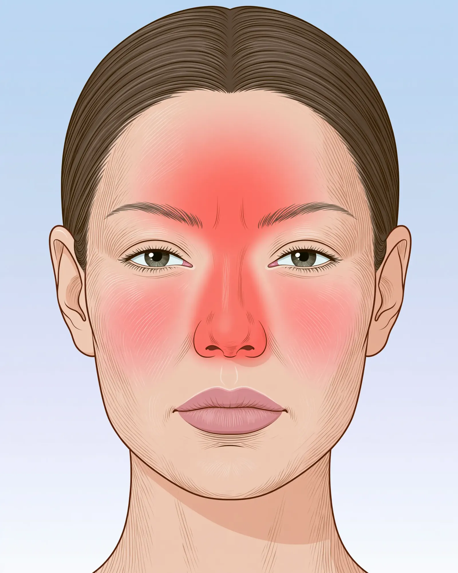
8. The Nose, Glabella, and Temples Deserve Special Attention to Vascular Risk
Although vascular complications can occur in many facial regions, certain areas warrant particular caution because of their vascular relationships and the potential severity of complications.
Clinical pearl: Anatomical risk should influence treatment selection before the procedure begins. The best technique cannot compensate for an inappropriate indication or inadequate emergency preparedness.
9. Skin Quality Is an Anatomical Variable, Not an Afterthought
Aesthetic outcomes depend on more than deep structures and facial volume. The skin itself plays a central role in how light reflects from the face, how contours appear, and how well tissues respond to treatment.
Epidermal pigmentation, dermal thickness, collagen organization, elastin, hydration, vascularity, and the effects of ultraviolet exposure all contribute to the patient's presentation.
Two patients with similar facial volume may have very different treatment needs because one has good elasticity and relatively uniform pigmentation, while the other has photodamage, laxity, textural irregularities, or pigmentary concerns.
This distinction is important when selecting among injectables, neuromodulators, lasers, radiofrequency, ultrasound-based treatments, and other modalities.
Skin tone and a history of post-inflammatory hyperpigmentation also influence treatment planning. The risk of pigmentary complications varies by procedure, device parameters, skin characteristics, and the underlying condition being treated.
Clinical pearl: Assess skin quality, pigmentary risk, and tissue laxity independently from volume loss. A comprehensive treatment plan may need to address several different anatomical problems.
10. Individualized Assessment Is More Important Than Memorizing Injection Maps
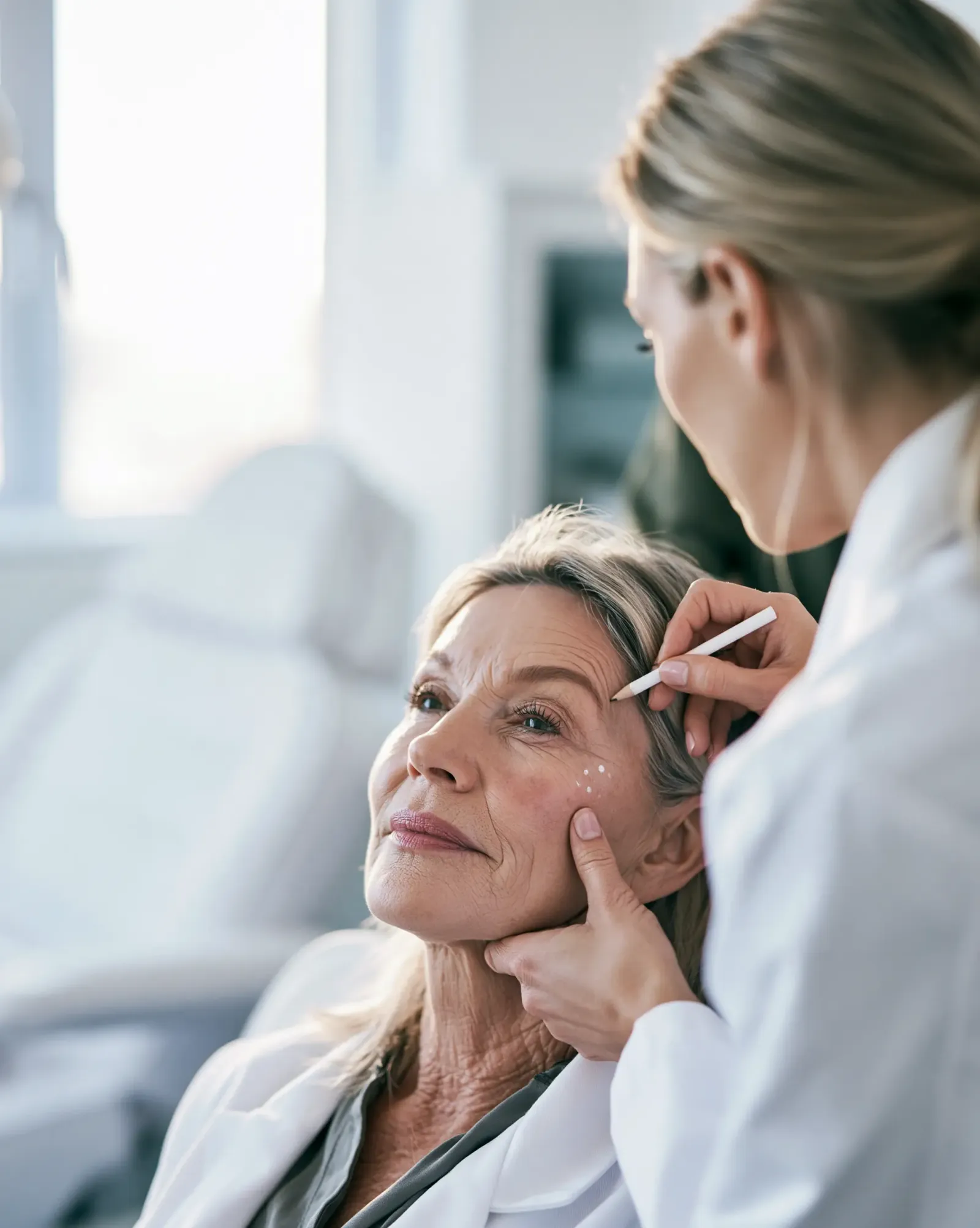
Documentation should reflect the assessment, treatment rationale, informed consent, and relevant risk discussion.
For clinicians who use ultrasound or other imaging to assess facial structures, these tools can provide additional anatomical information when used with appropriate training. However, imaging does not replace a sound understanding of anatomy or established safety protocols.
A successful aesthetic treatment is not defined by how much product is used or how many areas are treated. It is defined by whether the intervention appropriately addresses the patient's concern while respecting function, anatomy, safety, and individual goals.
Clinical pearl: The best injectors do not follow maps blindly. They use anatomical knowledge to interpret the patient in front of them.
Bringing It All Together
Facial anatomy is not a subject that can be mastered once and then set aside. It requires continuous study, observation, and refinement as clinical techniques and technologies evolve.
The 10 principles discussed here share a common message: successful aesthetic practice requires an understanding of how facial structures interact, how they change over time, and how interventions can influence both appearance and function.
For aesthetic healthcare professionals, this knowledge informs better patient selection, more thoughtful treatment planning, and more responsible management of complications.
The goal is not simply to produce a beautiful result. It is to achieve an appropriate, natural-looking outcome while preserving the patient's health, function, and individuality.
My advice to every aesthetic injector: Before you pick up the syringe, make sure you understand the anatomy. Your clinical judgment—and your patient's safety—depend on it.
Continue Your Education With Dr. Ellen Turner
At DrEllenTurner.com, my goal is to help aesthetic healthcare professionals strengthen their clinical foundations, evaluate emerging technologies critically, and apply evidence-informed principles to everyday practice.
Subscribe for weekly educational articles on facial anatomy, injectable treatments, dermatology, energy-based devices, regenerative medicine, and the business of aesthetic practice.